1. Biomechanical Foundations & Clinical Evolution of Intramedullary Nailing
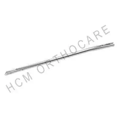
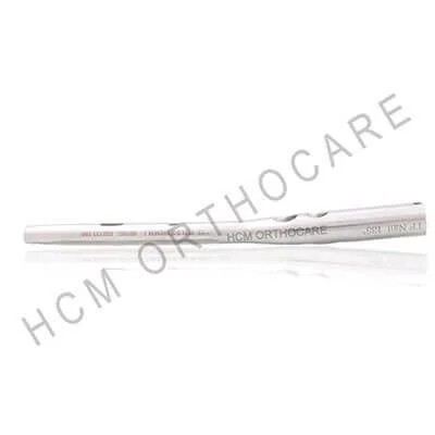
In modern trauma orthopedics, the treatment of long bone shaft fractures—specifically of the femur, tibia, and humerus—has undergone a paradigm shift from extramedullary plating to closed intramedullary interlocking nailing. As a primary internal load-sharing construct, an intramedullary interlocking nail is inserted directly into the medullary canal of a long bone. By aligning with the central neutral axis of the bone, the nail inherently minimizes the mechanical bending moments experienced under muscular contraction and weight-bearing forces.
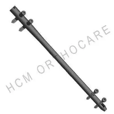
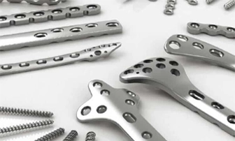
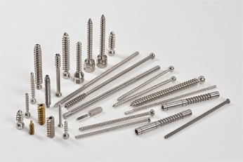
Unlike extramedullary bone plates, which act as load-bearing structures eccentric to the anatomical axis and are prone to mechanical fatigue failure, micro-motion disruption, and extensive soft-tissue stripping, intramedullary interlocking nails preserve the periosteal blood supply. The addition of proximal and distal locking screws (transfixion bolts) converts a simple intramedullary rod into a rigid rotational and axial stabilizing matrix. This biomechanical configuration successfully counteracts shear, torsional, and compressive loads across simple, segmental, and highly comminuted fracture patterns.
Information Gain: Neutral Axis Alignment vs. Eccentric Load Distribution
Positioning an implant along the intramedullary axis reduces structural bending moments by up to 60% compared to lateral eccentric plating. This load-sharing dynamic permits controlled axial micro-motion (0.2 mm to 1.0 mm), which stimulates osteoblast activity, accelerates secondary fracture healing via robust callus formation, and substantially reduces rates of non-union and implant fatigue failure.
Structural Fixation Comparison: Intramedullary Nailing vs. Alternative Modalities
To assist clinical procurement directors, trauma surgeons, and hospital inventory managers in evaluating fixation protocols, the table below details the mechanical and biological trade-offs of key orthopedic techniques:
| Evaluation Vector |
Intramedullary Interlocking Nails |
Extramedullary Locking Plates |
External Fixation Systems |
| Load Axis Distribution |
Central Neutral Axis (Load-Sharing) |
Eccentric Lateral Axis (Load-Bearing) |
External Axis (High Bending Moment) |
| Biological Envelope Impact |
Minimal disruption; preserves periosteum |
Moderate-to-high soft tissue periosteal stripping |
Percutaneous pin insertion; high pin-tract infection risk |
| Rotational & Torsional Control |
Superior via multi-planar locking screws |
Excellent via bi-cortical locking screws |
Moderate; reliant on pin frame geometry |
| Callus Formation & Bone Healing |
Secondary healing via osteogenic micro-motion |
Primary direct union (rigid compression) |
Secondary callus formation under controlled stress |
| Early Weight-Bearing Capability |
Immediate to early partial weight-bearing |
Delayed until radiographic bridging evidence |
Restricted due to pin-bone interface loosening |
| Implant Fatigue Failure Risk |
Extremely low due to coaxial stress alignment |
Moderate-to-high in non-union scenarios |
High pin loosening rate over prolonged periods |